
매달, 우리는 1000명 이상의 사람들이 시험 준비를 잘하고 시험을 잘 통과할 수 있도록 도와줍니다.
Abdomen Sonography Examination 온라인 연습
최종 업데이트 시간: 2025년10월10일
당신은 온라인 연습 문제를 통해 ARDMS AB-Abdomen 시험지식에 대해 자신이 어떻게 알고 있는지 파악한 후 시험 참가 신청 여부를 결정할 수 있다.
시험을 100% 합격하고 시험 준비 시간을 35% 절약하기를 바라며 AB-Abdomen 덤프 (최신 실제 시험 문제)를 사용 선택하여 현재 최신 165개의 시험 문제와 답을 포함하십시오.
정답:
Explanation:
The tunica albuginea is the dense fibrous capsule that directly surrounds the testicular parenchyma. The tunica vaginalis is a serous covering surrounding the testis externally, and the dartos fascia and pampiniform plexus are part of the scrotal wall and spermatic cord, respectively.
According to Rumack’s Diagnostic Ultrasound:
“The tunica albuginea is the fibrous capsule surrounding the testis and forming septa within the gland.”
Reference: Rumack CM, Wilson SR, Charboneau JW, Levine
D. Diagnostic Ultrasound. 5th ed. Elsevier, 2017.
AIUM Practice Parameter for Scrotal Ultrasound, 2020.
정답:
Explanation:
The renal arteries originate from the abdominal aorta just inferior to the superior mesenteric artery (SMA). The SMA arises anteriorly from the abdominal aorta at the level of L1, and just below it, the renal arteries branch laterally. The splenic vein, portal vein, and hepatic artery are located more superiorly in relation to the renal arteries.
According to Moore's Clinically Oriented Anatomy:
"The superior mesenteric artery arises from the anterior surface of the abdominal aorta just above the renal arteries." (Moore KL et al., Clinically Oriented Anatomy, 8th ed.)
Reference: Moore KL, Dalley AF, Agur AMR. Clinically Oriented Anatomy. 8th ed. Wolters Kluwer, 2018.
Gray’s Anatomy for Students, 4th ed., Elsevier, 2019.
정답:
Explanation:
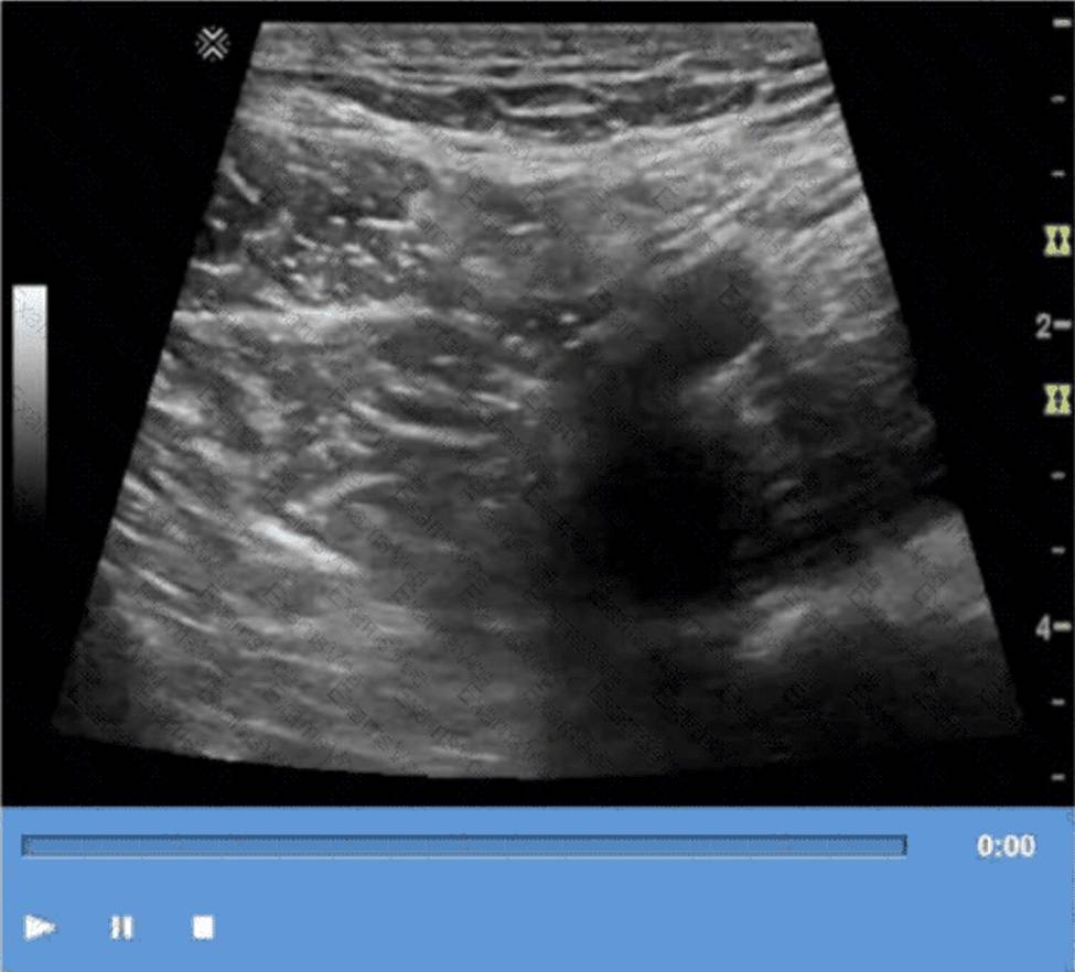
The technique shown in the video is compression. In ultrasound imaging―especially of soft tissue masses, the bowel, or venous structures―compression is used to evaluate the compressibility of structures. The image demonstrates a classic grayscale ultrasound view of a lesion or structure being compressed with the probe.
Compression sonography is particularly important in:
Evaluating venous patency (e.g., for deep vein thrombosis)
Differentiating cystic from solid structures
Evaluating bowel wall abnormalities or intussusception
Assessing lymph nodes and soft tissue masses (as shown here)
When a structure compresses easily under probe pressure, it suggests that the lesion is fluid-filled or soft. In contrast, incompressibility may indicate a solid mass or thrombus.
Differentiation from other options:
B. Valsalva: Involves forced expiration against a closed airway, used primarily to assess venous reflux or inguinal hernias―not what is demonstrated here.
C. Exhalation: A respiratory maneuver that passively alters thoracoabdominal pressure, not actively performed by the operator or causing focal structural change.
D. Deep inspiration: Used to improve visualization of the liver, diaphragm, or gallbladder―not to evaluate the compressibility of soft tissue.
References:
Rumack CM, Wilson SR, Charboneau JW, Levine
D. Diagnostic Ultrasound. 5th Edition. Elsevier, 2018.
Chapter: Ultrasound Technique and Physics, pp. 35C39.
AIUM Practice Parameter for the Performance of a Diagnostic Ultrasound Examination, 2020.
정답:
Explanation:
Von Hippel-Lindau (VHL) syndrome is a genetic disorder associated with multiple pancreatic cysts, pancreatic neuroendocrine tumors, and other systemic neoplasms. While cystic fibrosis can produce thickened pancreatic secretions, it rarely causes true pancreatic cysts.
According to Rumack’s Diagnostic Ultrasound:
“Multiple pancreatic cysts are strongly associated with Von Hippel Lindau syndrome.”
Reference: Rumack CM, Wilson SR, Charboneau JW, Levine
D. Diagnostic Ultrasound. 5th ed. Elsevier, 2017.
WHO Classification of Digestive System Tumors, 5th ed., IARC, 2019.
정답:
Explanation:
Hepatic adenomas are benign liver tumors strongly associated with long-term use of oral contraceptives and are most frequently found in women under 40. Hepatoblastoma is seen in children; hepatoma (HCC) is a malignant tumor typically found in cirrhotic livers. Cavernous hemangioma is unrelated to oral contraceptives.
According to Rumack’s Diagnostic Ultrasound:
“Hepatic adenomas occur predominantly in young women with a history of oral contraceptive use.”
Reference: Rumack CM, Wilson SR, Charboneau JW, Levine
D. Diagnostic Ultrasound. 5th ed. Elsevier, 2017.
WHO Classification of Tumours of the Digestive System, 5th ed., IARC, 2019.
정답:
Explanation:
Adrenal adenomas are the most common adrenal tumors in adults. They are often discovered incidentally (adrenal incidentalomas) and are usually nonfunctioning, though some may secrete cortisol or aldosterone. Neuroblastoma is common in children, pheochromocytomas are rarer catecholamine-producing tumors, and adrenal cortical carcinoma is malignant but much less common than adenomas.
According to Rumack’s Diagnostic Ultrasound:
“Adrenal adenomas are the most common adrenal masses in adults, frequently identified incidentally on imaging studies.”
Reference: Rumack CM, Wilson SR, Charboneau JW, Levine
D. Diagnostic Ultrasound. 5th ed. Elsevier, 2017.
ACR Incidental Findings Committee Guidelines for Adrenal Masses, 2017.
정답:
Explanation:
Urinomas are collections of urine in the retroperitoneum that result from trauma, surgery, or obstruction causing urine leakage. Trauma is a frequent cause of urinoma formation due to disruption of the urinary tract.
According to Rumack’s Diagnostic Ultrasound:
“Urinomas may develop as a complication of trauma, surgery, or obstructive uropathy with urinary extravasation into the retroperitoneum.”
Reference: Rumack CM, Wilson SR, Charboneau JW, Levine
D. Diagnostic Ultrasound. 5th ed. Elsevier, 2017.
AIUM Practice Parameter for Renal Ultrasound, 2020.
정답:
Explanation:
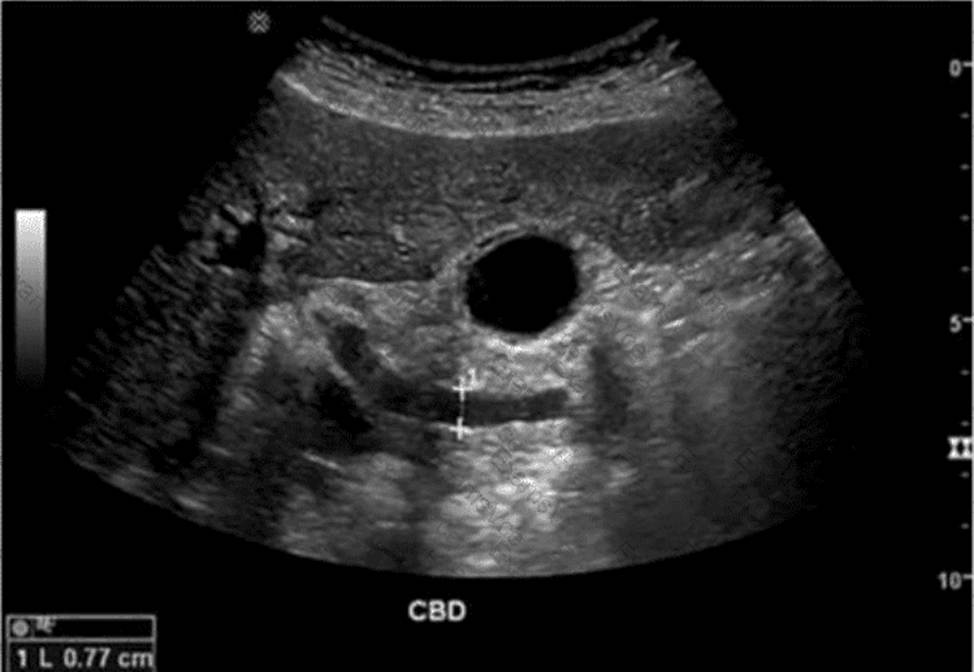
The ultrasound image demonstrates a dilated common bile duct (CBD), measuring approximately 7.7 mm in diameter. A normal CBD should generally measure less than 6 mm in a patient under 60 years old and may increase approximately 1 mm per decade thereafter or after cholecystectomy.
In the absence of gallstones within the CBD, one of the most concerning causes of CBD dilation is distal obstruction due to an extrinsic compressive lesion. The most common and clinically significant cause of distal CBD obstruction is a mass at the head of the pancreas.
A pancreatic head mass (e.g., adenocarcinoma) may compress the distal CBD and pancreatic duct simultaneously, resulting in the “double duct sign” ― dilation of both the CBD and pancreatic duct. This is a classic finding in pancreatic cancer.
Comparison of answer choices:
A. Liver mass ― unlikely to cause isolated CBD dilation unless invading the porta hepatis.
B. Cystic duct stone ― may cause gallbladder hydrops but typically not CBD dilation unless Mirizzi syndrome is present.
C. Pancreatic head mass ― Correct. This is the most likely cause of painless progressive CBD dilation without visible intraductal stones.
D. Gallbladder stones ― These may be associated with biliary colic or cholecystitis but typically do not cause CBD dilation unless the stone has migrated and obstructed the distal duct.
References:
Rumack CM, Wilson SR, Charboneau JW, Levine
D. Diagnostic Ultrasound, 5th ed. Elsevier; 2017.
Lee JK, Sagel SS, Stanley RJ.Computed Body Tomography with MRI Correlation, 4th ed. Lippincott Williams & Wilkins; 2006.
ACR Appropriateness Criteria® Right Upper Quadrant Pain (2021).
정답:
Explanation:
Transitional cell carcinoma (TCC) typically presents as a non-mobile, polypoidal, focal intraluminal mass projecting from the bladder wall. Mobility of the lesion helps differentiate TCC from blood clots or debris.
According to Rumack’s Diagnostic Ultrasound:
“Bladder TCC most often appears as a non-mobile, polypoid mass attached to the bladder wall.”
Reference: Rumack CM, Wilson SR, Charboneau JW, Levine
D. Diagnostic Ultrasound. 5th ed. Elsevier, 2017.
AIUM Practice Parameter for Bladder Ultrasound, 2020.
정답:
Explanation:
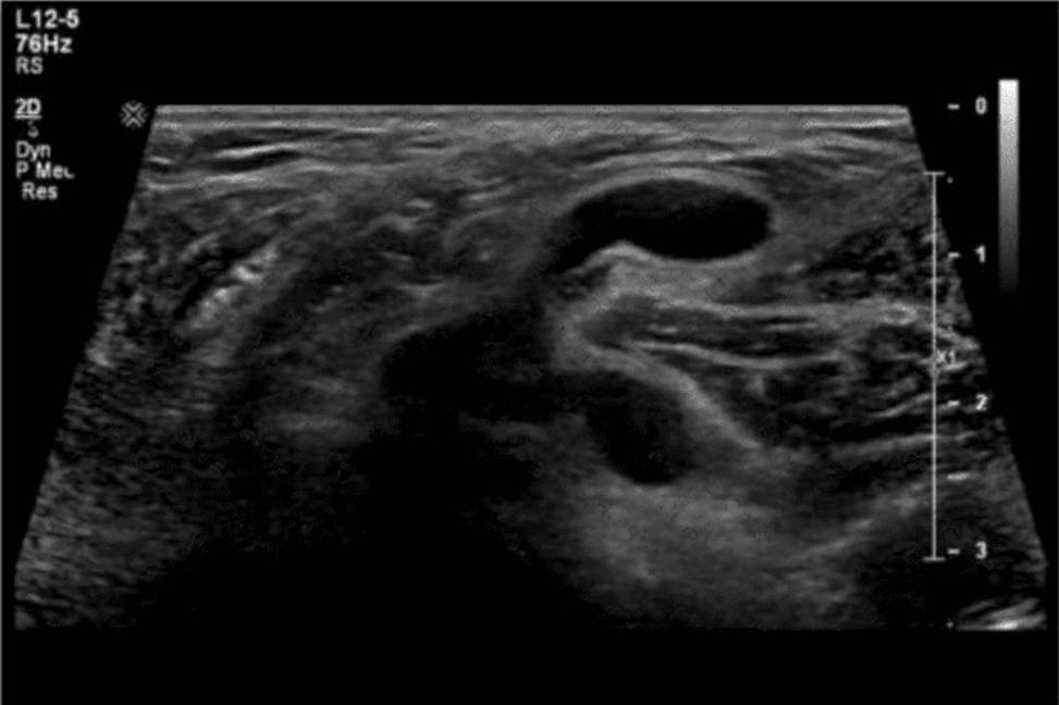
The ultrasound image demonstrates bowel loops with peristalsis visualized within the inguinal canal, which is diagnostic of an inguinal hernia―more specifically, an indirect inguinal hernia. Indirect hernias pass through the deep inguinal ring and may extend into the scrotum, appearing sonographically as bowel-containing masses adjacent to or within the scrotal sac. Peristaltic motion confirms the presence of viable bowel content.
This finding is typical in indirect inguinal hernias, which are more common in males and often congenital due to a patent processus vaginalis. The herniated bowel can be traced through the inguinal canal, as seen in this image.
Comparison of answer choices:
A. Hematocele presents as a complex fluid collection surrounding the testis, often due to trauma―no complex fluid or trauma is apparent here.
B. Testicular rupture shows discontinuity of the tunica albuginea and irregular testicular contour―none of which is seen.
C. Orchiectomy would show an absent testis―this is not the case here.
D. Indirect hernia is correct. The presence of bowel with peristalsis in the inguinal canal is diagnostic.
References:
Rumack CM, Wilson SR, Charboneau JW, Levine
D. Diagnostic Ultrasound, 5th ed. Elsevier; 2017.
AIUM Practice Parameter for the Performance of Scrotal Ultrasound Examinations (2021).
Dogra VS, Gottlieb RH, Rubens DJ, Oka M. Sonography of the scrotum. Radiology. 2003;227(1):18C36
정답:
Explanation:
An abdominal aortic aneurysm (AAA) measuring #5.5 cm represents a significantly increased risk of rupture and often requires surgical evaluation. The sonographer must report this critical finding immediately to the interpreting physician. The sonographer should never disclose a diagnosis directly to the patient.
According to AIUM and SRU Guidelines:
“An aortic diameter of 5.5 cm or greater should be promptly reported to the interpreting physician due to the high risk of rupture.”
Reference: AIUM Practice Parameter for Abdominal Aortic Ultrasound, 2020.
Society of Radiologists in Ultrasound (SRU) Consensus Statement, 2003.
정답:
Explanation:
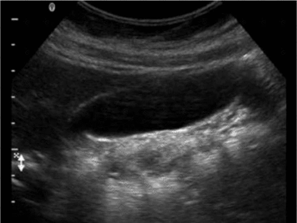
The ultrasound image demonstrates a fluid-filled structure in the posterior knee region, consistent with a Baker cyst (also called a popliteal cyst). A Baker cyst is a synovial fluid-filled sac arising from the posterior medial aspect of the knee joint, typically extending between the medial head of the gastrocnemius and the semimembranosus tendon.
The history of delayed-onset claudication (pain in the calf when walking) two weeks after this image was obtained is strongly suggestive of a ruptured Baker cyst. When a Baker cyst ruptures, synovial fluid may track inferiorly into the calf, producing pain, swelling, and clinical symptoms that mimic deep vein thrombosis (DVT) or arterial insufficiency (e.g., pseudothrombophlebitis syndrome).
Ultrasound findings consistent with a ruptured Baker cyst:
Complex fluid collection tracking along muscle fascial planes (hypoechoic to anechoic)
Posterior calf swelling and tenderness
Absence of thrombus in the deep venous system
Crescent-shaped fluid may be seen between muscle compartments
Why the other choices are incorrect:
A. Neuropathy: Would not show fluid-filled structures on ultrasound and would not present with calf swelling.
B. Infected hematoma: May appear complex, but would require a history of trauma or anticoagulation and systemic signs (fever, redness).
C. Thrombophlebitis: Involves a thrombosed superficial vein with wall thickening and surrounding inflammation, which is not seen in this image.
References:
American Institute of Ultrasound in Medicine (AIUM). Practice Guidelines for Musculoskeletal Ultrasound Examination, 2020.
Bianchi S., Martinoli
C. Ultrasound of the Musculoskeletal System. Springer, 2007. Chapter: Knee Region ― Popliteal Fossa and Baker’s Cyst, pp. 433C437.
Radiopaedia.org. Ruptured Baker cyst: https://radiopaedia.org/articles/ruptured-bakers-cyst
정답:
Explanation:
The ultrasound image demonstrates multiple parallel echogenic lines within the gallbladder lumen, extending distally and diminishing in intensity. These equidistant lines are classic for a reverberation artifact.
Reverberation occurs when the ultrasound beam reflects multiple times between two strong reflectors (such as the anterior gallbladder wall and the transducer), creating repeating echoes that appear as equally spaced lines.
This is frequently seen in:
The gallbladder (especially when collapsed or near the wall)
Near air-filled structures
With metallic objects or surgical clips
Reverberation does not represent a true anatomical structure and should be distinguished from real pathology.
Comparison of answer choices:
A. Banding refers to speed displacement or slice-thickness artifacts and typically does not appear as repetitive lines.
B. Shadowing is a dark band posterior to highly attenuating structures like gallstones or bone, not present here.
C. Attenuation is a general term for the reduction in sound energy as it travels―posterior acoustic shadowing is one type.
D. Reverberation ― Correct. The characteristic bright, parallel lines within the gallbladder lumen confirm this artifact.
References:
Kremkau FW. Sonography Principles and Instruments, 9th ed. Elsevier; 2015.
Rumack CM, Wilson SR, Charboneau JW, Levine
D. Diagnostic Ultrasound, 5th ed. Elsevier; 2017.
Hagen-Ansert SL. Textbook of Diagnostic Sonography, 8th ed. Elsevier; 2017.
정답:
Explanation:
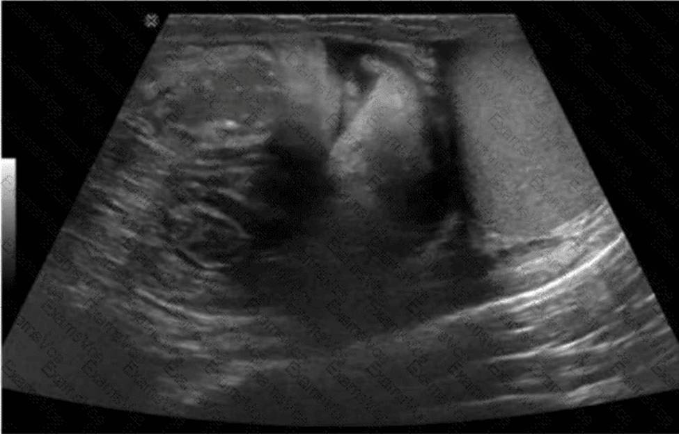
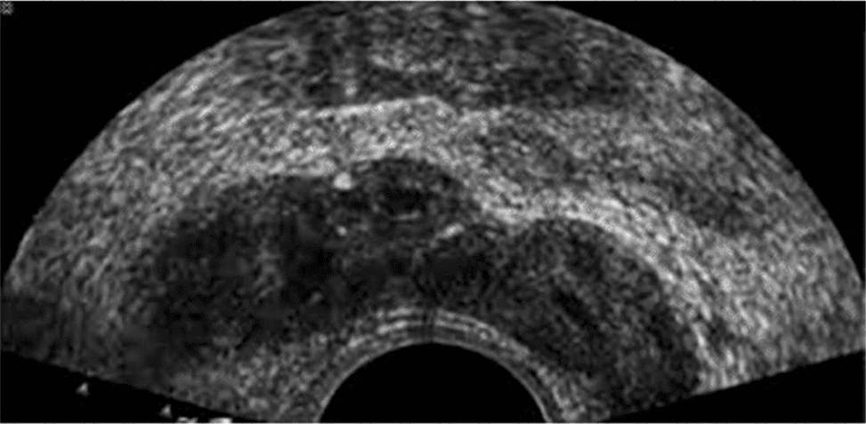
The ultrasound image shown is a transverse endorectal (transrectal) ultrasound, commonly used to evaluate the prostate and adjacent structures. The two hypoechoic (dark) oval-shaped structures seen superior and posterior to the prostate are characteristic of the seminal vesicles.
The seminal vesicles are paired, elongated glands located superior and posterior to the base of the prostate and are best visualized in transverse planes on endorectal imaging. They appear as hypoechoic or anechoic structures with internal septations, depending on the degree of fluid content.
In contrast:
The urethra appears as a central echogenic linear structure within the prostate.
The prostate base is more inferior in the scan and is visualized just above the urethra.
The ejaculatory ducts are usually not as prominently visualized and are located medial to the seminal vesicles, entering the prostate near the verumontanum.
This image most clearly demonstrates the bilateral seminal vesicles.
References:
Rumack CM, Wilson SR, Charboneau JW, Levine
D. Diagnostic Ultrasound, 5th ed. Elsevier; 2017.
ACRCAIUMCSRU Practice Parameter for the Performance of an Ultrasound Examination of the Prostate (2021).
Hagen-Ansert SL. Textbook of Diagnostic Sonography, 8th ed. Elsevier; 2017.
정답:
Explanation:
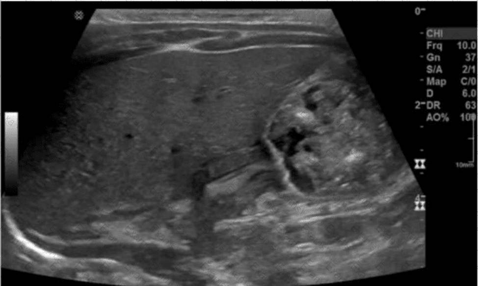
The ultrasound image demonstrates findings consistent with hypertrophic pyloric stenosis (HPS). This condition typically affects infants between 2 and 8 weeks of age and presents clinically with non-bilious projectile vomiting, weight loss, and dehydration.
In the ultrasound image, the classic “target” or “donut” sign can be seen in the transverse view of the hypertrophied pyloric muscle.
Key sonographic criteria for HPS include:
Pyloric muscle thickness #3 mm
Pyloric channel length #15C18 mm
This imaging appearance strongly correlates with the clinical presentation of projectile vomiting (Choice D), which is the hallmark symptom of HPS.
Comparison of answer choices:
A. Abnormal prenatal ultrasound (Choice A) is not typically associated with HPS, which develops postnatally.
B. Neonatal hyperbilirubinemia (Choice B) is not an indication for a pyloric ultrasound and affects liver /biliary imaging.
C. Red currant jelly stools (Choice C) are indicative of intussusception, not HPS.
D. Projectile vomiting (Choice D) is the most common clinical indication leading to an ultrasound exam that reveals HPS.
References:
Rumack CM, Wilson SR, Charboneau JW, Levine
D. Diagnostic Ultrasound, 5th ed. Elsevier; 2017.
AIUM Practice Parameter for the Performance of Ultrasound of the Pyloric Region in Infants (2014).
Hernanz-Schulman M. Infantile hypertrophic pyloric stenosis. Radiology. 2003;227(2):319C331.